When you hear “surgery,” you probably picture a long incision, a night in the hospital, and weeks of hobbling. Foot and ankle surgery has moved a long way from that image. Over the last decade, orthopedic and podiatric teams have adopted minimally invasive techniques that use tiny portals, specialized burrs and cameras, and fluoroscopic guidance to correct many common problems. The goal is straightforward: less soft-tissue disruption, less pain, smaller scars, and a quicker return to the shoes and activities that make up your daily life.
I have spent years in operating rooms and clinics watching these methods evolve. Done well, minimally invasive approaches can be transformative. Done on the wrong problem, or by the wrong hands, they can disappoint. This guide will help you understand what minimally invasive foot and ankle surgery is, when it makes sense, what recovery looks like, and how to choose a foot and ankle surgeon you trust.
What “minimally invasive” really means for the foot and ankle
Minimally invasive surgery, sometimes called percutaneous or keyhole surgery, uses incisions typically measured in millimeters instead of centimeters. In the foot and ankle, these portals allow a foot and ankle surgeon to work through the skin and soft tissue with slender instruments, often guided by live X-ray (fluoroscopy) or a small camera. Compared with traditional open surgery, the technique preserves more blood supply to skin and muscle, which is one reason patients often report less swelling and stiffness.
The other key difference is how bone is cut and reshaped. In an open bunion correction, for example, an orthopaedic foot and ankle surgeon might make a several-centimeter incision over the big toe joint, then use a saw to perform an osteotomy and fix it with screws. In a minimally invasive bunion surgery, the surgeon uses a 2 to 4 millimeter burr through a tiny incision to make the same cuts under fluoroscopy, then stabilizes the correction with small screws placed through puncture sites. The mechanics are similar, but the soft-tissue handling is very different.
Not every foot and ankle problem is a candidate for this approach. Deep infections, complex fractures with multiple fragments, severe deformities requiring extensive realignment, and some arthritic joints are still best treated through open techniques. An experienced foot and ankle doctor will explain where minimally invasive methods help and where they do not.
Conditions commonly treated through small incisions
Sports injuries, bunions, hammertoes, heel pain, ankle instability, and cartilage damage make up a large portion of what we treat. Many, though not all, can be managed with minimally invasive foot and ankle surgery.
Bunions and forefoot deformities. Minimally invasive bunion surgery has matured quickly. Modern techniques can correct mild to many moderate bunions with percutaneous osteotomies secured by screws. Hammertoes may be corrected through tiny portals with tendon releases and bone remodeling. The benefits patients notice most are less swelling at the big toe and fewer problems fitting into a shoe early in recovery. That said, large bunions with first metatarsal instability may need a Lapidus fusion, which can be performed through a smaller open approach or a hybrid technique. A foot and ankle bunion surgeon should tailor the plan to your anatomy and goals rather than forcing a one-size method.
Heel pain and plantar fasciitis. When months of measured treatment fail, a partial plantar fascia release can be done endoscopically through two small incisions. The goal is to alleviate tension while preserving arch stability. The majority of people with plantar fasciitis never need surgery, but for the stubborn cases, a surgeon for plantar fasciitis can explain the pros and cons of an endoscopic release versus continued nonoperative care.
Achilles tendon problems. For acute Achilles tendon ruptures, percutaneous repair with small incisions and suture-passing devices can reduce wound complications, which matter when the skin behind the ankle is tight. For chronic tendinopathy, minimally invasive debridement and calcaneal spur shaving can reduce pain while preserving the healthy tendon. Not every rupture is a candidate for a small-incision repair. High-level athletes, complex tear patterns, or significant tendon degeneration sometimes benefit from a more open approach with grafting. A surgeon for Achilles tendon injuries should walk you through those nuances.
Ankle instability and ligament injuries. Many lateral ligament reconstructions combine arthroscopy with mini-open techniques to repair or augment the ligaments that hold the ankle steady. Arthroscopy allows the ankle arthroscopy surgeon to treat coexisting cartilage lesions and impingement at the same sitting. Patients with generalized ligament laxity or repeat sprains may need added reinforcement such as an internal brace. The decision is individualized after exam and imaging.
Cartilage damage and impingement. Ankle and foot arthroscopy through 2 to 4 millimeter portals lets a foot and ankle cartilage surgeon remove scar tissue, smooth cartilage flaps, perform microfracture for small defects, and address anterior ankle pinching. For midfoot and big toe joint arthritis, minimally invasive debridement can help, but advanced arthritis often requires fusion or joint replacement. Here again, an orthopedic surgeon for ankle pain will balance short-term relief against long-term function.
Fractures. Select fractures can be stabilized with percutaneous screws placed through tiny incisions. An ankle fracture with simple fibula and medial malleolus breaks might be amenable to limited incisions, but a high-energy pilon fracture with joint shattering is not. A foot and ankle fracture surgeon or foot trauma surgeon will use CT scans to plan whether a minimally invasive strategy is safe and stable enough to allow early motion.
Flatfoot and cavus foot corrections. Some realignments, such as calcaneal osteotomies, now have percutaneous options using burrs and screws, which can reduce wound complications in areas with limited soft tissue. Other components, like tendon transfers or spring ligament reconstruction, still require targeted open work. A foot deformity surgeon often blends techniques, choosing minimally invasive where it helps and open where needed for precision.
Joint fusion and replacement. Fusions of midfoot joints and the big toe joint remain open or mini-open in most practices, since joint preparation and fixation demand direct control. An ankle replacement surgeon typically uses a standard approach with careful soft-tissue handling rather than fully percutaneous methods, though navigation and patient-specific instruments have improved accuracy and reduced soft-tissue trauma. A foot and ankle joint replacement surgeon will advise you if joint preservation, fusion, or replacement fits best.
What to expect before surgery
A thoughtful evaluation sets the stage for a good result. Your foot and ankle physician will start with a careful history and physical exam, then use X-rays for alignment and arthritis, and sometimes MRI or weight-bearing CT for cartilage and subtle deformity. Expect a conversation about your activity level, work demands, shoes, and tolerance for a staged recovery. Minimally invasive techniques can shorten downtime, but bone still heals at its own pace. If a bunion osteotomy is performed percutaneously, it needs weeks to consolidate just like an open cut would.
In the clinic, I try to sketch the plan. If we are correcting a bunion, we map where small incisions will be, how the bone will nearby foot surgery options be shifted, and whether we will add a soft-tissue procedure to balance the joint. For ankle arthroscopy, we discuss portal sites, what cartilage work may be done, and whether a ligament repair is likely based on exam and imaging. Clear expectations reduce anxiety and improve adherence to the plan.
Anesthesia matters as much as the incisions. Most minimally invasive foot and ankle surgeries are done as outpatient procedures under a regional block plus light sedation. A popliteal or saphenous nerve block numbs the limb for hours after surgery, which cuts down on early pain medication. If you have sleep apnea or other conditions, the anesthesiologist adjusts the plan. Good communication between the foot and ankle surgical specialist and anesthesia team is essential.
Inside the operating room, in plain language
Perioperative details can feel opaque. Here is the rhythm. After a safety check, the orthopedic foot and ankle surgeon marks your foot and uses fluoroscopy to verify alignment. Small incisions are made, each protected with a sleeve so the burr or shaver does not heat the skin. Saline often flows to cool the instruments and carry away debris. For bone cuts, the surgeon advances the burr under live X-ray until the desired line is created, then shifts the bone using clamps and fixes it with screws placed through tiny stab incisions. If we are scoping a joint, we inflate the joint with fluid and insert a pencil-sized camera to visualize cartilage, ligaments, and synovium.
The tech runs the fluoroscopy unit to provide two or three planes of view. The surgical nurse tracks instruments and implants. Times vary by procedure. A percutaneous bunion can take 45 to 90 minutes depending on severity. An ankle arthroscopy with ligament repair may run 60 to 120 minutes. Precision matters more than speed. The smaller the incision, the more reliant we are on imaging and tactile feedback to ensure perfect cuts and screw trajectories.
Recovery, realistically
Marketing tends to promise “back on your feet in no time.” It is better to think in phases. Minimally invasive incisions heal quickly, but bone and ligaments do not fast-forward healing just because the skin cut is small. Patients often report less pain and swelling, which helps them mobilize earlier and avoid stiffness, but the timeline depends on the procedure.
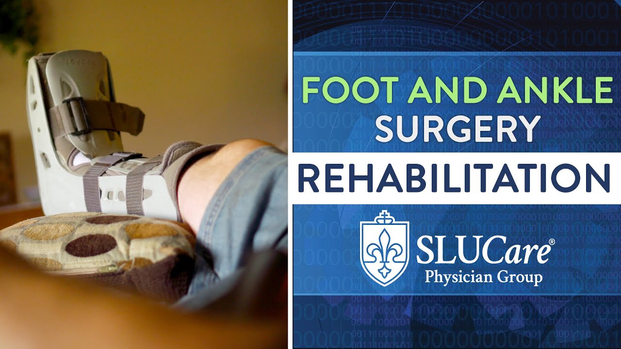
Swelling control is your first job. Elevation, compression, and a measured walking plan are key. Many patients can bear weight immediately in a protected boot after percutaneous forefoot procedures. Some ankle arthroscopy patients are full weight bearing within days. Ligament reconstructions and osteotomies may require weeks of protected load. Your foot and ankle care specialist will tailor a schedule that balances healing with preventing joint stiffness.
Physical therapy starts early in many cases. Gentle range of motion keeps joints supple. Scar care, even for tiny scars, matters. I stress shoe choices at each milestone. Wider toe boxes for bunion work, heel counters for instability, and rocker soles for big toe stiffness can make recovery smoother. Most desk workers return in 1 to 2 weeks with a boot under the desk. Jobs that require standing or lifting take longer, often 4 to 8 weeks. Runners typically resume light jogging around 8 to 12 weeks after cartilage debridement or bunion correction if healing looks good, later if bone was cut or ligaments reconstructed.
Pain management has changed. With regional anesthesia and local injections, most patients use a short course of anti-inflammatories and a limited number of opioid tablets, often fewer than ten. Ice, elevation, and a realistic plan reduce the need for more.
Risks and trade-offs you should hear up front
No surgery is risk-free, even through small incisions. The odds of wound complications and infection are lower with minimally invasive approaches, particularly in areas where the skin is thin. On the other hand, working through small portals can increase the risk of incomplete correction or malpositioned screws if the surgeon does not have the right equipment or experience. Radiation exposure is higher for the surgical team due to reliance on fluoroscopy, which is why your orthopedic surgeon for foot injuries wears protective gear and keeps exposures short.
Nerve irritation can occur, especially superficial sensory nerves around the ankle and toes. It is usually temporary but can linger. For bunions, the challenge is balancing correction with joint motion. Overcorrection can lead to stiffness or hallux varus, undercorrection to recurrence. For percutaneous Achilles repair, the sural nerve runs close to the surgical field and must be respected. A fellowship trained foot and ankle surgeon or a podiatric surgeon with advanced training will be candid about these details and your personal risk profile.
Another trade-off is tactile control. In open surgery, you see and feel every structure. In percutaneous surgery, you visualize under X-ray and feel through instruments. It requires a different skill set. The learning curve is real. Ask how your surgeon trained and how many of your exact procedure they perform annually.
Who is a good candidate, and who is not
You are more likely to benefit if your goals match what minimally invasive techniques deliver: pain relief, correction of mild to moderate deformity, smaller scars, and a faster early recovery. You also need to be reliable with postoperative instructions. Small incisions do not give you a free pass to ignore weight-bearing limits.
Certain conditions push us toward open or Springfield, NJ foot and ankle surgeon hybrid surgery. Severe bunions with first ray instability, advanced ankle arthritis, complex fractures, diabetic Charcot deformity, and revisions with significant scar tissue often need more exposure. If you smoke, your risk of wound problems and bone healing delays rises. Good surgeons will be frank about waiting to operate until you have optimized your health, sometimes working with your primary doctor to control diabetes or help you reduce nicotine use.
Age alone rarely rules you out. I have seen healthy seventy-year-olds recover better than sedentary forty-year-olds. What matters is bone quality, circulation, skin condition, and your support system at home. A foot and ankle clinic with a multidisciplinary team can help you prepare, from prehab to postoperative home setups.
Choosing the right foot and ankle specialist
Titles vary. You might meet an orthopedic foot and ankle surgeon, an orthopaedic foot and ankle specialist, or a podiatry surgeon. What counts is focused training and experience. Fellowship training in foot and ankle surgery, whether through orthopedic or podiatric pathways, means an extra year devoted to complex cases and advanced techniques. Board certification demonstrates a standardized level of knowledge and peer-reviewed practice. Look for a board certified foot and ankle surgeon who performs your exact procedure regularly and can show you typical outcomes and complication rates.
Ask practical questions. How many minimally invasive bunion corrections did you perform last year? What percentage required conversion to an open approach? For ankle arthroscopy with ligament stabilization, what is your re-sprain rate at one year? If a complication occurs, how do you handle it? Good surgeons welcome informed questions. Reviews can help you gauge communication and office flow, but the consult is where you judge trust and clarity. A top rated foot and ankle surgeon is the one who makes you feel heard, explains options without pressure, and has the skills to execute the plan safely.
Cost, insurance, and the logistics no one tells you
Minimally invasive does not necessarily mean more expensive. Many procedures are outpatient, which lowers facility costs. Insurance coverage depends on medical necessity, not incision size. Your orthopedic doctor for foot and ankle conditions will document failed conservative care and functional limitations. Implant choice can affect price. Some low-profile screws are costlier than standard hardware, but the difference is usually small compared with facility and anesthesia fees. If you have a high-deductible plan, ask the scheduler for a preauthorization and an estimate. Clarify what is included: surgeon fee, facility, anesthesia, implants, postoperative boot, and follow-up visits.
Logistically, plan your first two weeks as if you had a bigger surgery. Line up rides, a shower stool, a clear path at home, and meals ready to go. Even with tiny incisions, you will move better and heal faster if the basics are handled.
My take on specific procedures patients ask about
Minimally invasive bunion correction. For the right bunion, it delivers. Expect earlier shoe comfort and less stiffness compared with larger open approaches. A foot corrective surgeon will measure angles on weight-bearing X-rays and examine first ray stability to decide if percutaneous osteotomies are enough or if you need a fusion closer to the arch.
Endoscopic plantar fascia release. It is a last resort after diligent stretching, night splints, shockwave therapy, and injections. When needed, a limited release can free you from morning pain. Over-releasing can flatten the arch, so the surgeon for heel pain should be conservative and precise.
Percutaneous Achilles repair. I use it for midsubstance ruptures in active patients who want faster rehab and a smaller scar, provided the gap is not excessive. We protect the sural nerve and test the repair in the operating room. Expect a boot with wedges and early controlled motion. Return to running often starts around 4 to 5 months, cutting at 6 to 9 months.
Ankle arthroscopy with ligament repair. It is an excellent option for recurrent sprains with mechanical instability. Benefits include treating cartilage lesions at the same time and smaller incisions for ligament work. Physical therapy is crucial. Most athletes return to sport between 3 and 6 months depending on the demands.
Percutaneous calcaneal osteotomy for flatfoot reconstruction. The smaller incision reduces wound issues in a tight area, but the rest of the reconstruction may still be open. I view this as a hybrid, not purely minimally invasive, and I counsel patients accordingly.
Rehabilitation that respects biology
Regardless of technique, the principles of rehab do not change. The first two weeks focus on protecting the repair, controlling swelling, and maintaining mobility in joints that were not operated on. Weeks three to six generally introduce more motion and partial weight bearing or transitions from boot to shoe with support. By weeks six to twelve, bone cuts have consolidated enough to load more normally, proprioception work picks up, and balance training returns. Athletes start linear drills, then cutting and plyometrics when single-leg strength and landing mechanics look good.
A sports foot and ankle surgeon will coordinate closely with your therapist and coach if you are a competitive athlete. The goal is not just getting back, but lowering the odds of reinjury. For chronic ankle instability, for example, your risk of re-sprain drops if your peroneal strength and balance exceed your pre-injury baselines before you return.
When minimally invasive is not the right choice
It bears repeating. If you have severe arthritis with bone-on-bone contact and deformity, a joint-sparing minimally invasive procedure may offer short-lived relief. A foot and ankle arthritis surgeon might steer you toward a fusion or, for the ankle, a replacement. For complex fractures with joint involvement, an ankle reconstruction surgeon may need open reduction to restore anatomy precisely. Infections require exposure and debridement. Diabetic Charcot collapse needs stability above all else, often through robust hardware and larger exposures. The best foot and ankle orthopedists do not chase tiny incisions at the expense of long-term function.
A brief story that shows the difference
A distance runner in her forties came to the foot and ankle clinic with a moderate bunion and pain after ten miles. Orthotics and shoe changes had helped for a while. On exam, her first metatarsal was stable, and imaging showed a hallux valgus angle in the mid 20s. She wanted to return to half marathons without chronic pain. We planned a minimally invasive bunion correction with percutaneous osteotomy and two low-profile screws.
She was in a boot for four weeks, started range of motion right away, and transitioned to a stiff-soled shoe at week five. At eight weeks, she jogged short intervals on a track. By four months, she ran her usual 10 miles without the blistering and redness she used to notice over the bump. Could she have done well with an open approach? Likely. Did the smaller incisions and reduced swelling help her stay engaged with rehab and return sooner? In her case, yes.
How to prepare yourself to be a great surgical partner
- Clarify your goals in writing. Pain relief for daily life is different from returning to cutting sports or long hikes, and it guides the plan. Ask your foot and ankle surgical specialist to outline the first six weeks day by day, including weight bearing, wound care, and work restrictions. Line up a recovery space at home, with elevation options and clear paths to the bathroom and kitchen. Commit to smoking cessation and glucose control if applicable, at least six weeks before and after surgery. Choose shoes in advance for transitional phases, such as a wide, supportive sneaker for forefoot surgery or a heel-stable shoe for ankle procedures.
The bottom line patients remember
Minimally invasive foot and ankle surgery is not a magic trick. It is a set of refined tools and techniques in the hands of a specialist who understands anatomy, biomechanics, and healing. When chosen thoughtfully, it reduces soft-tissue trauma and often speeds early recovery without compromising the quality of correction. The best outcomes come from matching the right operation to the right problem, then executing the rehab plan with the same care as the operation itself.
Whether you work with an orthopedic podiatric foot surgeon, a sports injury foot and ankle surgeon, or an orthopaedic foot and ankle surgeon, look for experience, transparency, and a plan that makes sense to you. Ask clear questions, prepare well, and respect the biology of healing. Small incisions can deliver big results, but only if the fundamentals are in place.